CT scan
Computed tomography (CT) uses X-rays taken from many angles and a computer to create detailed cross-sectional images. It is fast — especially useful in emergencies.
- Head injury and suspected stroke
- Chest, abdomen and pelvis problems
- Kidney stones
- Cancer detection, staging and follow-up
- Guiding biopsies
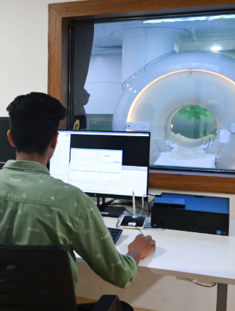
Smita Memorial Hospital has a Philips Incisive 128-slice CT scanner that delivers high-resolution images with reduced radiation exposure, and performs CT-guided biopsy.
MRI
Magnetic resonance imaging (MRI) uses a strong magnetic field and radio waves — no X-rays — to create very detailed images, particularly of soft tissues.
- Brain and spinal cord
- Disc prolapse and nerve compression
- Joints, ligaments and cartilage
- Prostate imaging
- Detailed evaluation of some tumours
Smita Memorial Hospital has a Philips 1.5T MRI.
Key differences
- CT is quicker; MRI takes longer
- CT uses X-ray radiation; MRI does not
- MRI shows soft tissues in greater detail
- MRI is noisier and involves lying in a narrower space
- Metal implants can affect MRI safety — always tell the team
Contrast dye
Some scans need a contrast agent injected into a vein to highlight blood vessels or abnormalities. Tell the team if you have kidney problems, diabetes medicines, or previous reactions to contrast.
Preparing for your scan
- Follow fasting instructions if given
- Tell staff about pregnancy
- For MRI, remove all metal and tell staff about pacemakers, implants, clips or metal fragments
- Bring previous scan reports and images
- Tell the team if you feel anxious in closed spaces
Results
Reports are prepared promptly by radiologists, who are available to discuss examinations with patients and doctors when needed. Radiology provides round-the-clock emergency support.
Other imaging tests you may hear about
- Digital X-ray — quick images of bones, chest and abdomen; Smita Memorial Hospital uses a Carestream DRX Ascend 630 mA system
- Ultrasound — uses sound waves, no radiation; ideal for abdomen, pregnancy, thyroid and soft tissues
- Doppler scan — ultrasound that shows blood flow in arteries and veins
- Mammography — low-dose X-ray of the breast
- Echocardiography — ultrasound of the heart
MRI and claustrophobia
Some people feel anxious inside an MRI scanner. Let the team know in advance. They can explain each step, keep in contact with you during the scan and give you a call button. Closing your eyes, breathing slowly and focusing on music or counting can help. Your doctor can discuss medicine for relaxation if needed.
Imaging in pregnancy and for children
Always tell the team if you are or might be pregnant. Doctors choose tests without radiation, such as ultrasound or MRI, where possible, and weigh benefits carefully before any X-ray or CT. For children, imaging is tailored to use the lowest necessary radiation, and a parent can usually stay nearby.
Image-guided procedures
Imaging is also used to guide procedures without open surgery. At Smita Memorial Hospital, radiologists perform USG-guided FNAC and biopsy and CT-guided biopsy, and the Interventional Radiology department performs a wide range of image-guided treatments.
Specialists you can consult


Frequently asked questions
Is MRI safe?
MRI does not use radiation. It is safe for most people, but metal implants and devices must be checked beforehand.
Is CT radiation harmful?
CT uses a controlled dose of radiation. Doctors request it when the benefit outweighs the small risk. Smita Hospital's scanner is designed for reduced radiation exposure.
Are CT and MRI available at night?
Radiology at Smita Memorial Hospital provides round-the-clock emergency support.
Do I need an appointment?
Walk-ins are welcome; appointments are recommended for specialised scans needing preparation.
Sources
- Radiodiagnosis, Radiology & Diagnostics, smitahospital.com